Posts
Skin Histology & Thin skin v/s Thick skin
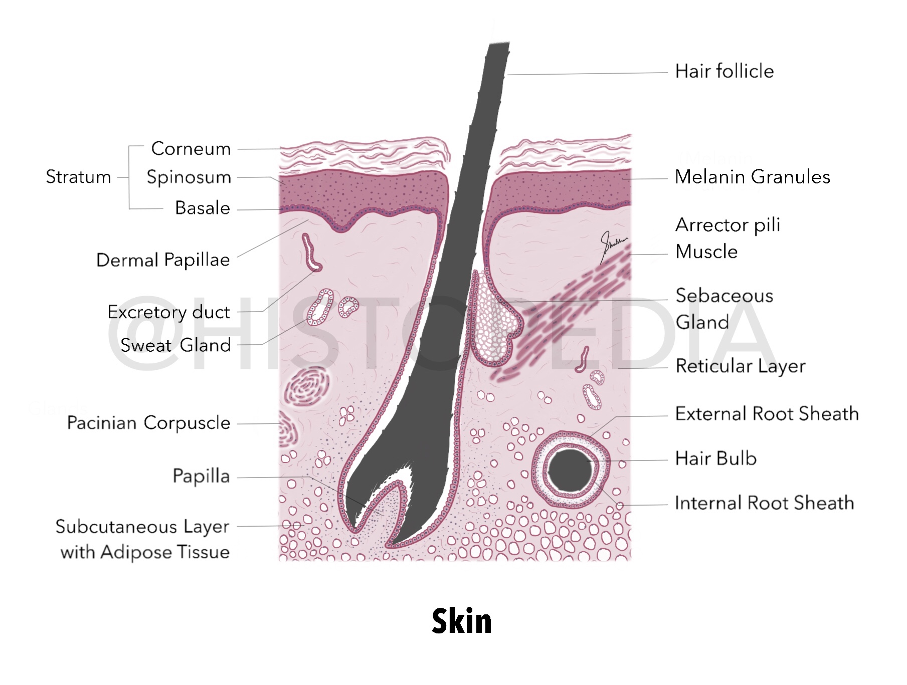
Layers: (top to bottom) Epidermis Stratum Corneum Stratum Spinosum Stratum Basale (with pigmented/melanin granules) Dermis Dermal Papillae (which forms our fingerprints) Reticular layer Subcutaneous layer with Adipose tissue Skeletal muscle fibers (not illustrated here) Hair follicle (numerous, closely packed and oriented at an angle to the surface) Hair bulb Internal root sheath External root sheath Connective tissue Sebaceous Glands : surround each hair follicle, aggregates of clear cells . Arrector pili muscle : smooth ms. aligned at an oblique angle to the hair follicle, it contracts to move hair follicle in more vertical position. Pacinian corpuscles : pressure and vibration sensing receptors. Other labels : sweat glands and blood vessels (omitted here) Imp. Difference between thin skin and thick skin Histology of both types is similar only difference is the thickness of epidermis . As skin of Palm and Soles are constantly exposed to wear/tear/abrasion, thicknes...
Dry/Compact Bone (T.S.)
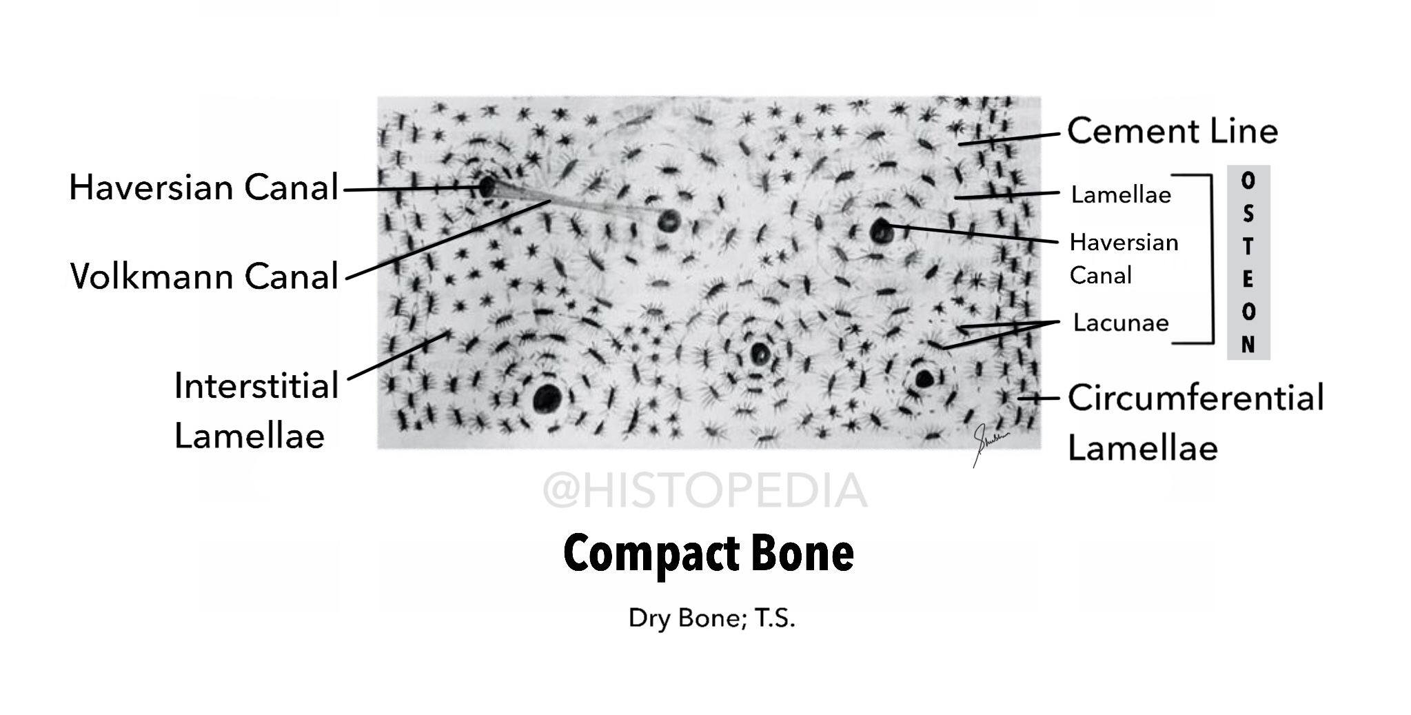
Grounded thin section of dried bone shows empty canals for blood vessels, lacunae for osteocytes and connecting canaliculi. Important Labels: Osteon ( Structural unit) Haversian Canal (central canal) Lamellae (thin plates of bone that contain Osteocytes) Lacunae (almond shaped spaces that contain Osteocytes) Cement line : boundary between each Osteon Volkmann Canal : also called Perforating Canal, it is anastomoses between central canals. External Circumferential lamellae : forms external wall of compact bone Internal Circumferential lamellae : forms internal wall of compact bone (endosteum along the marrow cavity). “In living bone lacunae of each Osteon house Osteocytes & Central/Haversian canals contain reticular connective tissue, blood vessels and nerves.”
Uterus (Follicular Phase)
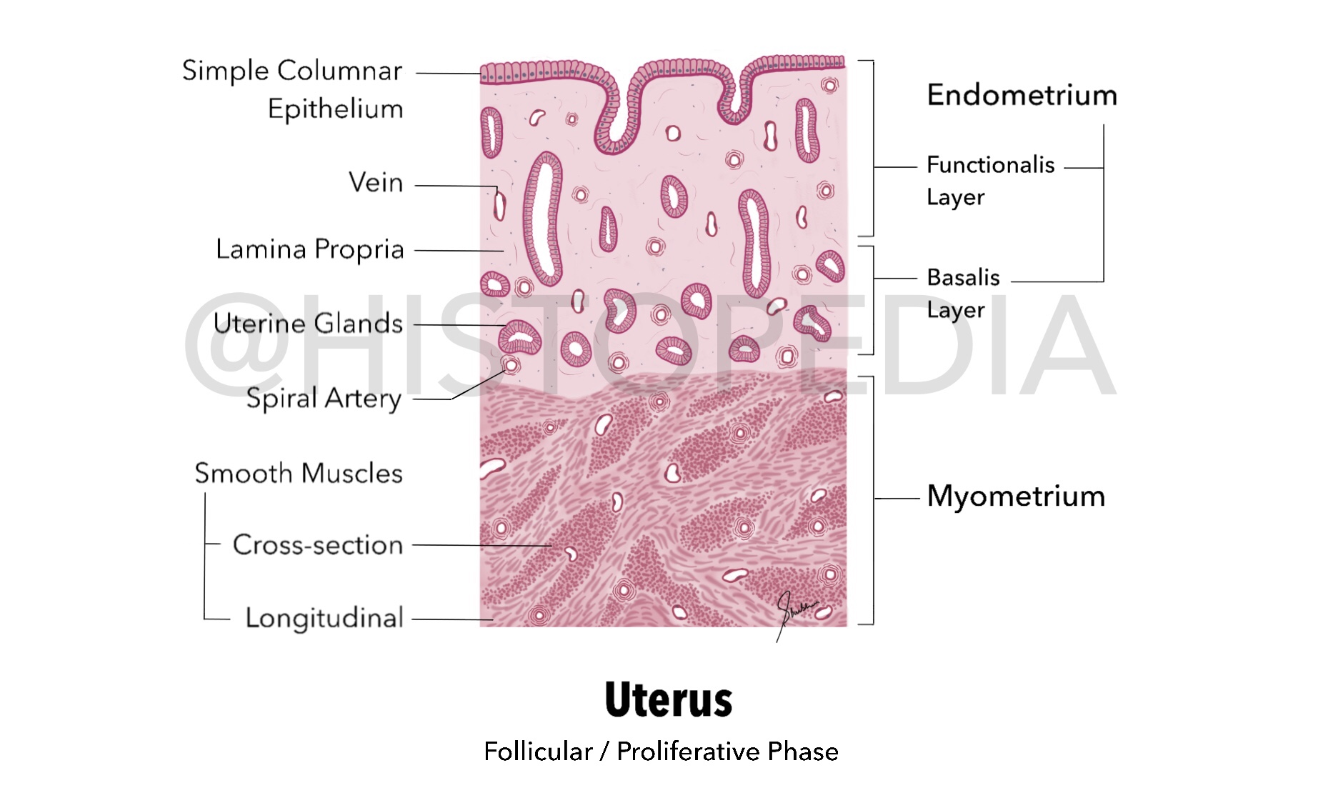
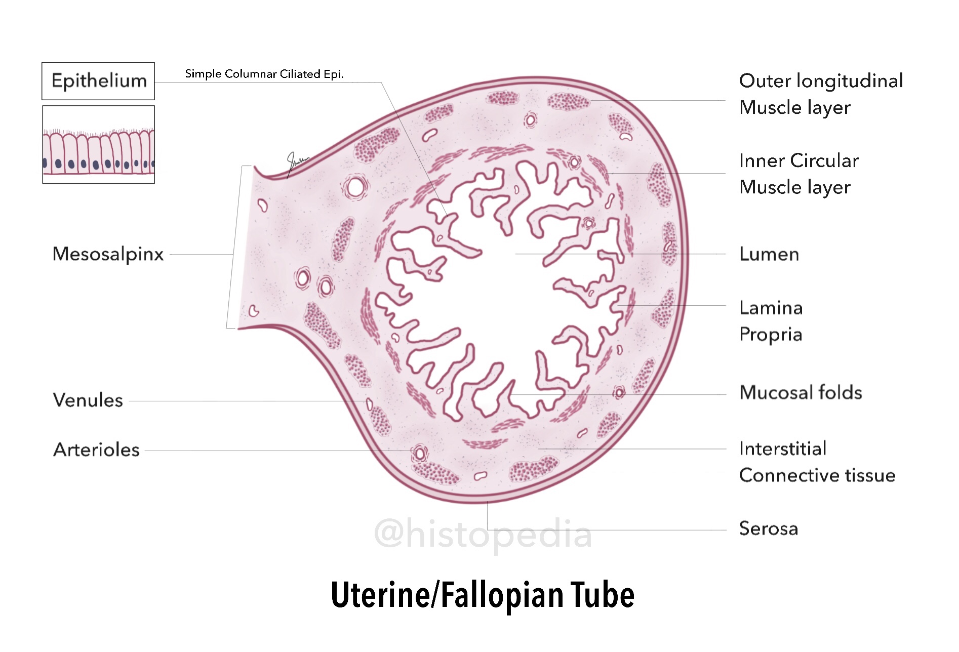
Surface lining of Endometrium is Simple Columnar Epithelium, which extends downwards forming long, tubular Uterine Glands. In Proliferative/Follicular Phase these glands are mainly straight in upper portion of endometrium and branch out in lower portion, because of which numerous glands are seen in cross section (in Basalis layer). During menstrual cycle, endometrium exhibit morphological changes that are correlated with ovarian functions. These cyclic changes are divided into Proliferative/follicular Phase Secretory/luteal Phase and Menstrual Phase Important Labels: Endometrium (Functionalis and Basalis Layers) Myometrium (Smooth longitudinal & cross-sectional muscles) Lining Epithelium Uterine Glands Spiral/Coiled Arteries
Lung (Panoramic View)

The histology of Intrapulmonary bronchi is similar to that of the trachea and extra-pulmonary bronchiole, except that C-shaped cartilage rings are absent here and replaced by cartilage plates. Intrapulmonary Bronchus : Pseudo-stratified Columnar Ciliated Bronchial Epithelium Cartilage Plates & Seromucous Bronchial Glands Terminal Bronchiole : Simple Columnar Epithelium Prominent mucosal folds & well developed smooth muscles Respiratory Bronchiole : Simple Cuboidal Epithelium Alveolar Out-pocketings in the wall Gives rise to alveolar ducts Alveolar Walls : Out-pocketings of respiratory bronchioles type 1 & 2 Pneumocyets Also contain Alveolar macrophages Bands of Smooth muscle fibres in interalveolar septa
Kidney
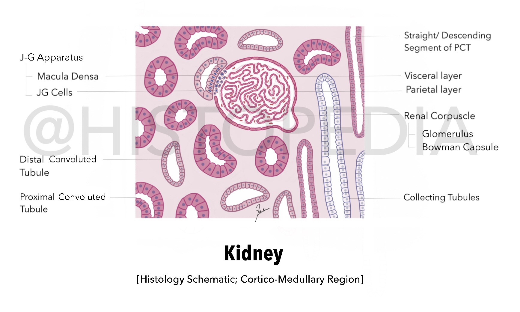
In sagittal section, the kidney shows outer dark staining cortex and inner light staining medulla. Externally cortex is covered with a dense irregular Renal Capsule. Important Labels: Proximal Convoluted Tubule Distal Convoluted Tubule Renal Corpuscle (Glomerulus & Bowman Capsule) J-G Apparatus Collecting tubules Identification Tip: Abundant Glomeruli (grainy appearance and dark staining) Elongated tubule like structures (lightly stained & adjacent to dark grainy region)
Large Intestine: Colon Wall (T.S.)

The Principal functions of large intestine are to absorb water and minerals/electrolytes from the remaining undigested material coming from small intestine, therefore the lining epithelium contains absorptive cells and numerous secreting cells (goblet cells) that lubricate the lumen. No digestive enzymes are produced. Characteristic Features/ Identification Points: Wall of Colon exhibit temporary folds, which consist of both Mucosa and Sub-mucosa. Villi are absent Lining Epithelium is characterized by Goblet Cells Intestinal Glands (Crypts of Lieberkühn) No Glands in Sub-mucosa Muscularis externa of the colon is Atypical, unlike small intestine. The Longitudinal Layer is arranged into strips or bands of smooth muscle called Taeniae Coli (3-bands).