Posts

Showing posts with the label Salivary Glands

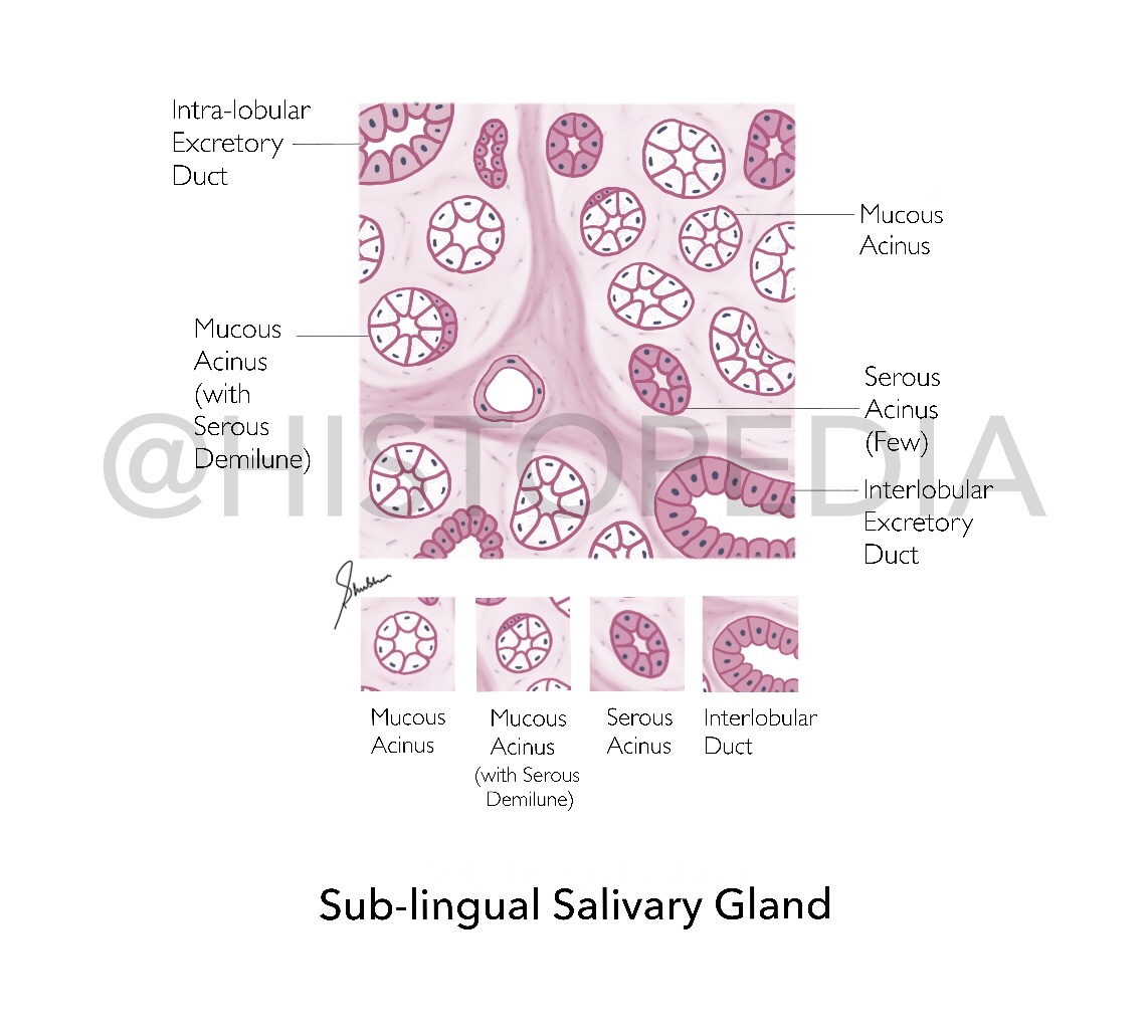
Sub-lingual Salivary Gland
Image
Identification Points:  1. Majority of acini are M ucous   2. Abundant  Serous Demilunes    [absent in Parotid Gland] 3. Intercalated ducts are short/absent (not readily observed) 4. Non-striated Intra-lobular ducts   [differentiating factor from other 2 salivary glands] 5. Abundant Interlobular Connective tissue septa   Description: It is a mixed type of salivary gland with both serous and mucous acini, most of which are mucous and capped with serous demilunes , It is responsible for 2-5% of saliva secretion which is mucoid in nature.  It is smallest salivary gland, almond shaped located in the floor of oral cavity on either side of tongue. Flow of Secretions: Mucoid Secretions  ⤵️ Non-striated Intra-lobular ducts  (equivalent to striated ducts of other 2 salivary glands)  ⤵️ Interlobular duct   ⤵️ 8-20 excretory ducts  (ducts of Rivinu) Step-by-step Illustration: File Diagram: How to differentiate betw...
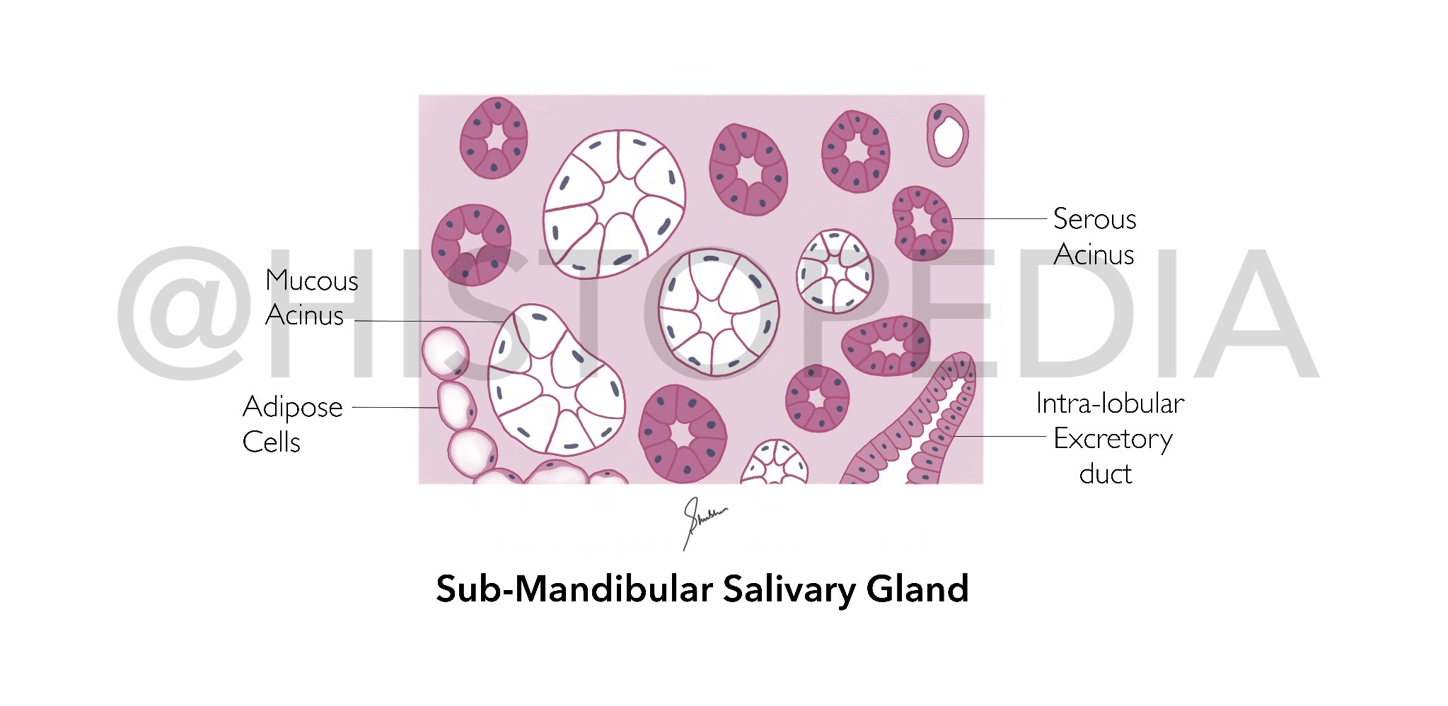
Sub-Mandibular Salivary Gland

Image
Identification Points:  1.   Mixed   acini   (both serous and mucous)   [differentiating factor b/w Submandibular and Parotid salivary gland] 2. Serous Demilunes    [absent in Parotid Gland] 3. Mucous acini are weakly stained (almost colourless cytoplasm)in contrast to serous acini  Description: It is a mixed type of salivary gland with both serous ( predominating ) and mucous acini, located below the Jaw and pour it’s secretions via Warhtin’s Duct in the floor of oral cavity in front of tongue. It is divided into deep and superficial parts by Myohyoid muscle. It is responsible for 70% of saliva secretion which is predominately mucoid in nature because mucous cells are more active. Flow of Secretions: Mucoid Secretions  ⤵️ Intercalated ducts   ⤵️ Striated Ducts (lined with  Simple Columnar Cells )  ⤵️ Intra-lobular ducts  ⤵️ Interlobular duct ( Pseudo-Stratified Columnar epithelium )  ⤵️ Warhtin’s Duct (fl...

Parotid Salivary Gland

Image
  Identification Points:  1.   Serous acini   (pyramid shaped secretory cells with round basal nucleus)  2. No Serous Demilune 3.  Striated Ducts Description: It is a large capsulated serous gland, subdivided into lobes and lobules by connective tissue septa located just anterior to the ears. It secretes 25% of total saliva which is serous (watery) in nature.  At higher magnifications small secretory granules are visible in apices of serous acini Flow of Secretions: Serous acini (secretions)  ⤵️ Intercalated ducts   ⤵️ Striated Ducts (lined with Simple Columnar Cells )  ⤵️ Intra-lobular ducts  ⤵️ Interlobular duct ( Pseudo-Stratified Columnar epithelium )  ⤵️ Stensen’s Duct (open near 2nd upper molar) Step-by-step Illustration: File Diagram: How to differentiate between all 3 Salivary Glands?

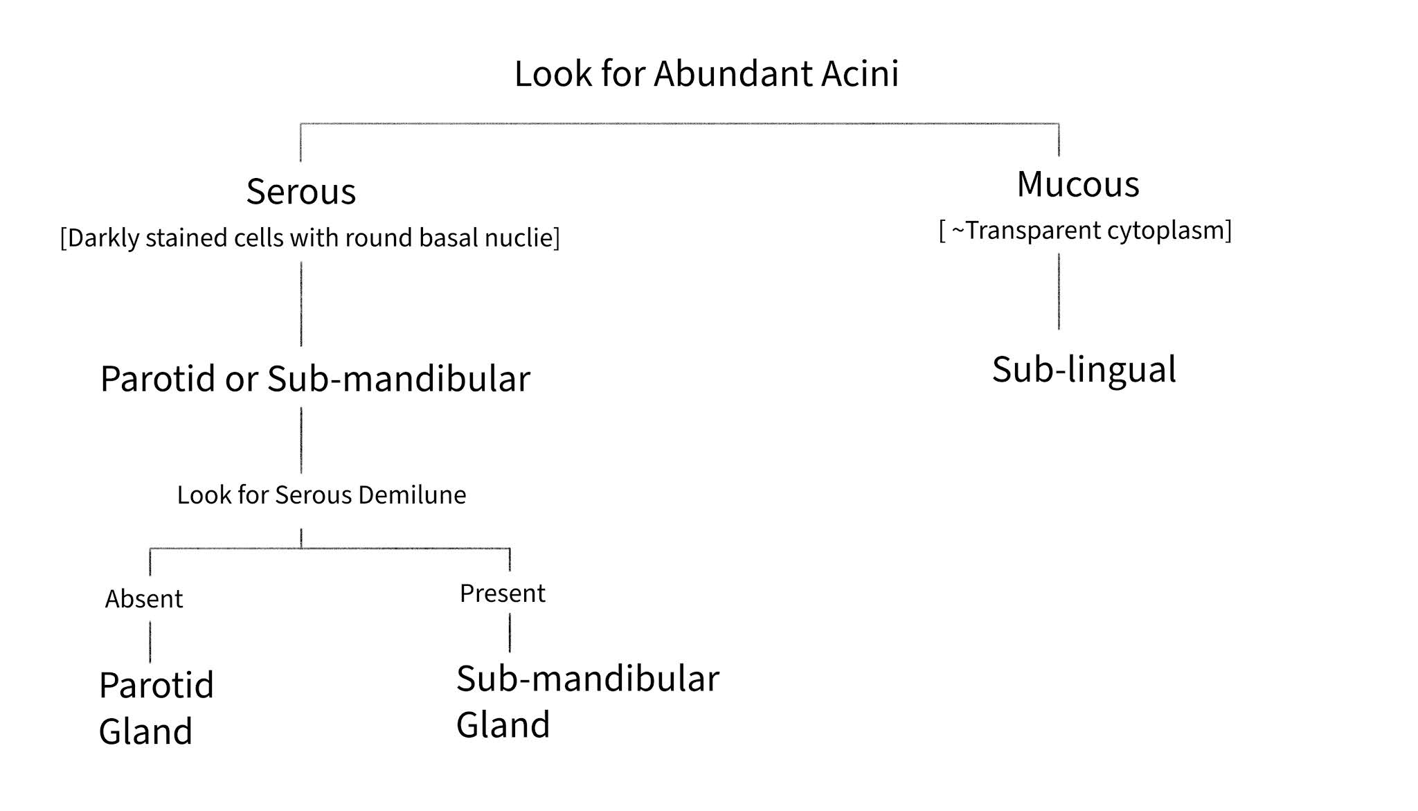
How to differentiate between all 3 Salivary Glands?

Image