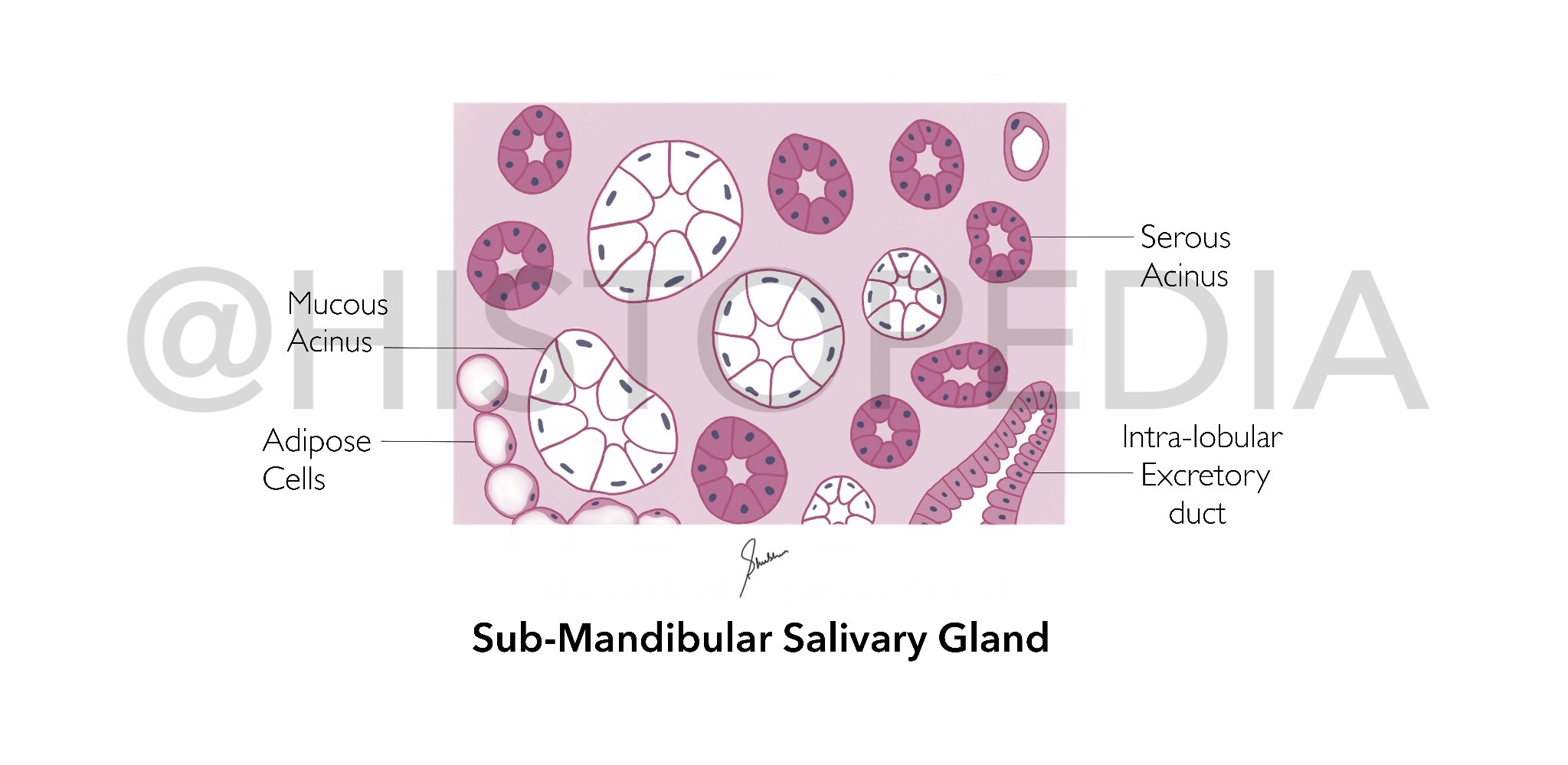
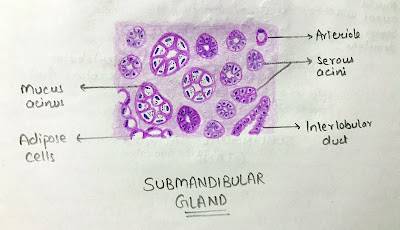
Sub-Mandibular Salivary Gland
Identification Points:
1. Mixed acini (both serous and mucous)
[differentiating factor b/w Submandibular and Parotid salivary gland]
2. Serous Demilunes
[absent in Parotid Gland]
3. Mucous acini are weakly stained (almost colourless cytoplasm)in contrast to serous acini
3. Mucous acini are weakly stained (almost colourless cytoplasm)in contrast to serous acini
Description:
It is a mixed type of salivary gland with both serous (predominating) and mucous acini, located below the Jaw and pour it’s secretions via Warhtin’s Duct in the floor of oral cavity in front of tongue. It is divided into deep and superficial parts by Myohyoid muscle.
It is responsible for 70% of saliva secretion which is predominately mucoid in nature because mucous cells are more active.
Flow of Secretions:
⤵️
Intercalated ducts
Intercalated ducts
⤵️
Intra-lobular ducts
Intra-lobular ducts
⤵️
Warhtin’s Duct (floor of oral cavity)


