Posts

Showing posts with the label Gastro Intestinal Tract (GIT)

Large Intestine: Colon Wall (T.S.)

Image
The Principal functions of large intestine are to absorb water and minerals/electrolytes from the remaining undigested material coming from small intestine, therefore the lining epithelium contains absorptive cells and numerous secreting cells (goblet cells) that lubricate the lumen. No digestive enzymes are produced.   Characteristic Features/ Identification Points: Wall of Colon exhibit temporary folds, which consist of both Mucosa and Sub-mucosa. Villi are absent Lining Epithelium is characterized by Goblet Cells  Intestinal Glands (Crypts of Lieberkühn)  No Glands in Sub-mucosa Muscularis externa of the colon is Atypical, unlike small intestine. The Longitudinal Layer is arranged into strips or bands of smooth muscle called Taeniae Coli (3-bands).

Stomach (Fundus & Body) T.S.

Image
Fundus and Body constitutes most extensive region of Stomach. There are 4 general regions ( inside to outside ) : Mucosa (Epithelium + Lamina Propria + Muscularis Interna) Submucosa Muscularis Externa (Circular & Longitudinal Smooth Muscle layers) Serosa Important Labels ( Additional to above 4 ) Parietal Cells &  Chief Cells (Gastric Glands) Gastric Pits Oblique Muscle Layer (Characteristic Feature)

Small Intestine: Duodenum (Longitudinal Section)

Image
.  

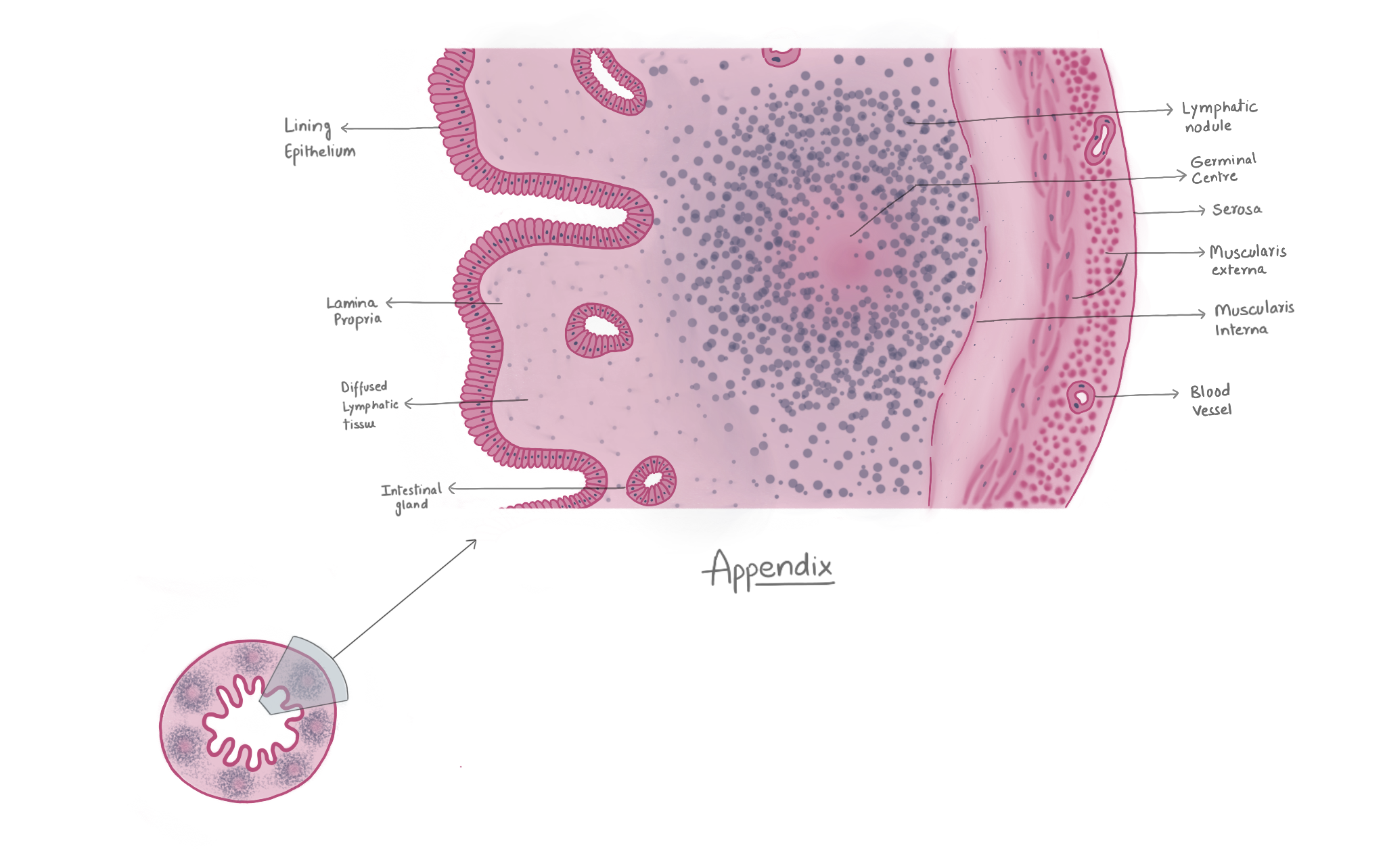
Appendix

Image
Characteristic Feature:   Presence of lymphoid tissue mass in mucosa and submucosa, similar to Peyers Patches of Intestine. It’s morphology is similar to that of Colon with some modifications like muscularis externa forms a continuous coat. Less developed intestinal glands. Abundant diffused lymphatic tissue. Important labels: Lymphatic Nodules (may extend to the submucosa) Mocosa Lining Epithelium ( Simple Columnar ) with Goblet Cells Intestinal Glands (Crypts of Lieberkühn)  Lamina Propria Submucosa (rich in blood vessels) Muscularis Mucosa (myenteric plexus between inner and outer layers) Serosa (with underlying adipose tissue)   

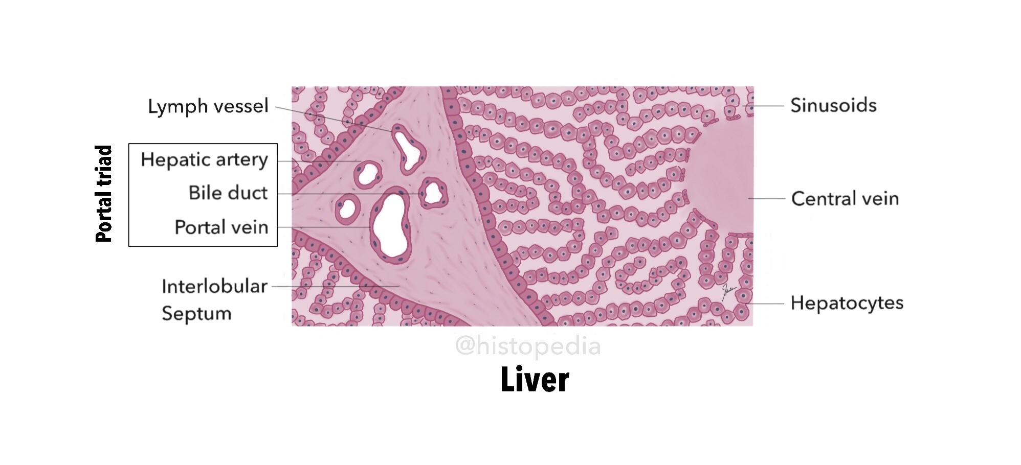
Liver (Hepatic Lobule and Portal Triad)

Image

Pancreas (Exocrine and Endocrine Portions)

Image
 

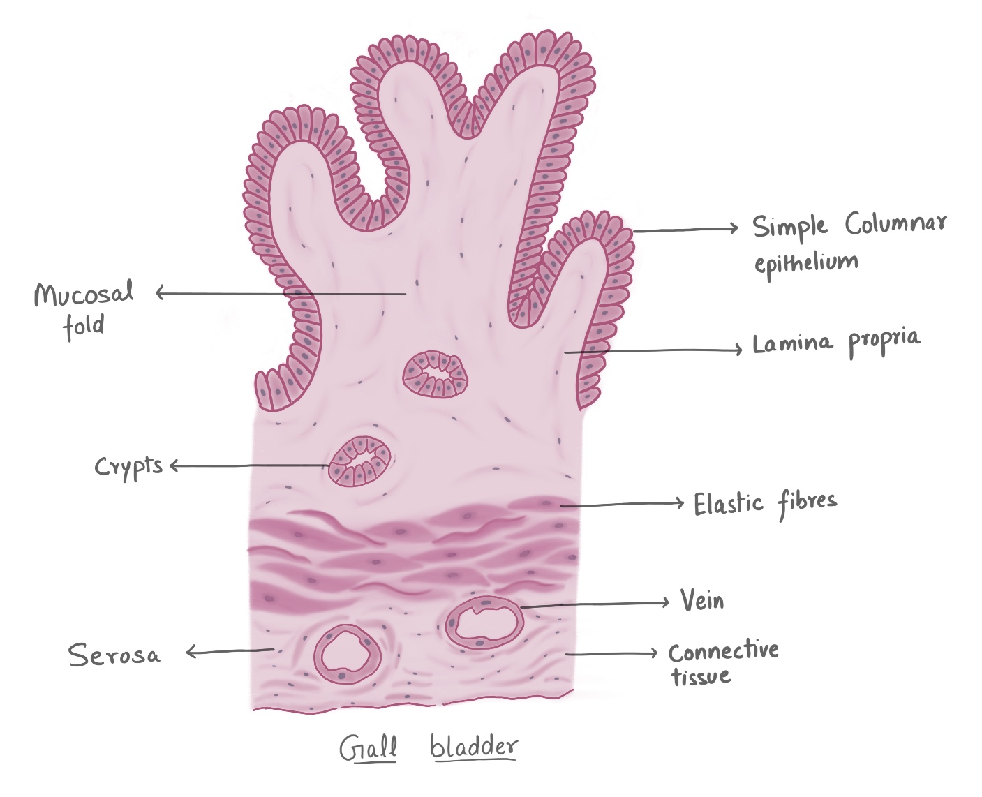
Gall Bladder

Image
Important Point: Sub-mucosa and Muscularis Mucosae are absent in Gall Bladder.  

Lower Esophagus

Image
  Serosa is only present in lower/ terminal esophagus & absent in the rest of the portion. Steps: