Posts

Showing posts with the label Connective tissue

Skin Histology & Thin skin v/s Thick skin
Image
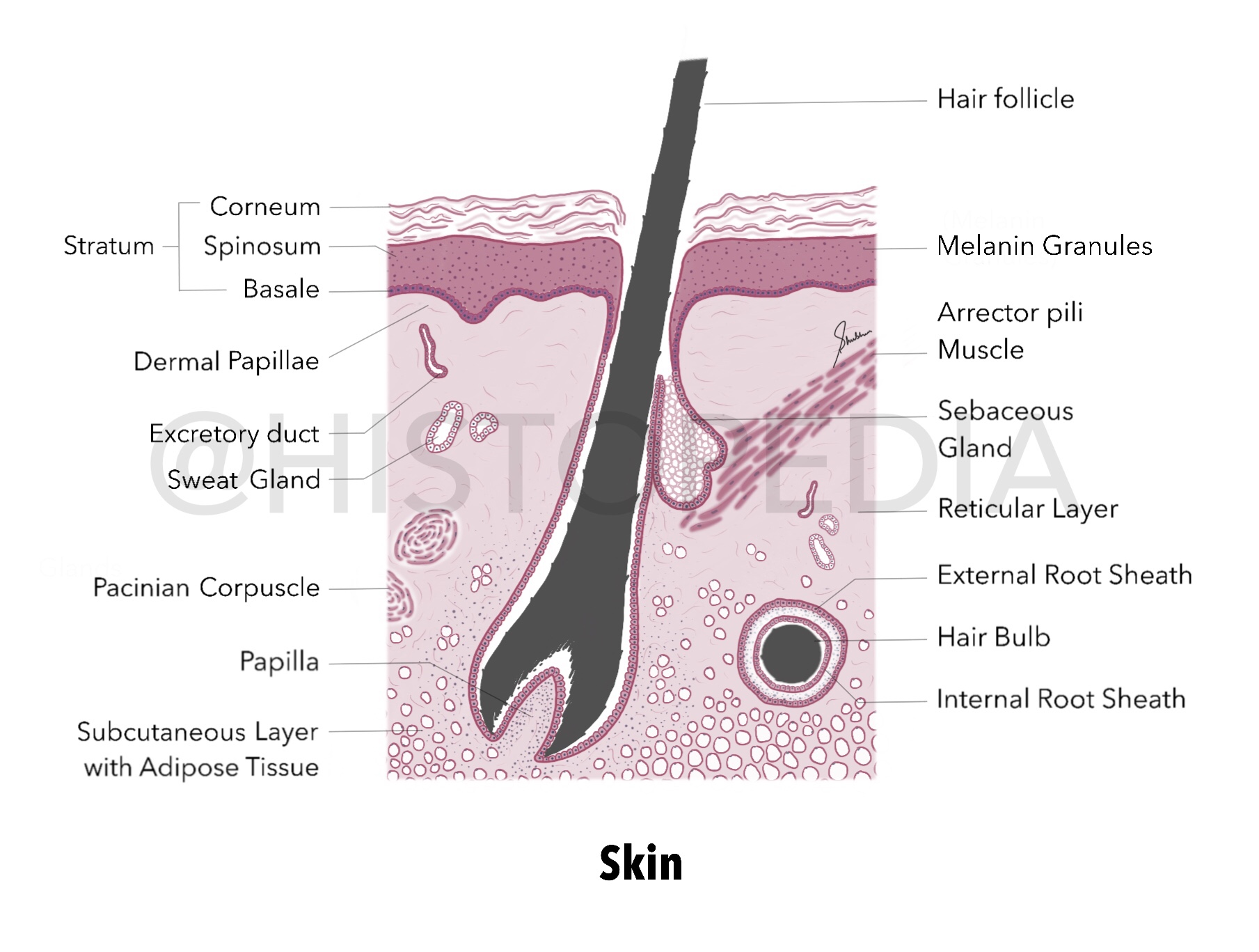
Layers: (top to bottom) Epidermis Stratum Corneum Stratum Spinosum Stratum Basale (with pigmented/melanin granules)  Dermis Dermal Papillae (which forms our fingerprints) Reticular layer Subcutaneous layer with Adipose tissue Skeletal muscle fibers (not illustrated here) Hair follicle (numerous, closely packed and oriented at an angle to the surface) Hair bulb Internal root sheath External root sheath Connective tissue Sebaceous Glands :  surround each hair follicle, aggregates of clear cells . Arrector pili muscle : smooth ms. aligned at an oblique angle to the hair follicle, it contracts to move hair follicle in more vertical position. Pacinian corpuscles : pressure and vibration sensing receptors. Other labels : sweat glands and blood vessels (omitted here) Imp. Difference between thin skin and thick skin   Histology of both types is similar only difference is the thickness of epidermis . As skin of Palm and Soles are constantly exposed to wear/tear/abrasion, thicknes...
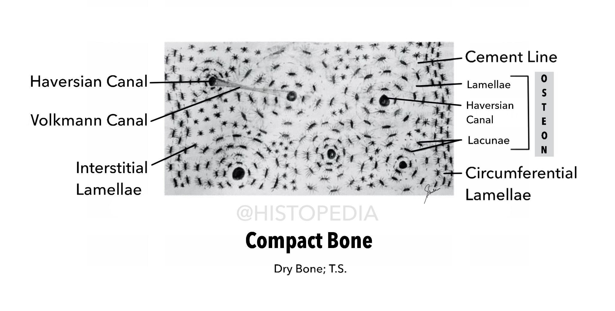
Dry/Compact Bone (T.S.)

Image
Grounded thin section of dried bone shows empty canals for blood vessels, lacunae for osteocytes and connecting canaliculi. Important Labels: Osteon ( Structural unit) Haversian Canal (central canal) Lamellae (thin plates of bone that contain Osteocytes) Lacunae (almond shaped spaces that contain Osteocytes) Cement line : boundary between each Osteon Volkmann Canal : also called Perforating Canal, it is anastomoses between central canals. External Circumferential lamellae : forms external wall of compact bone Internal Circumferential lamellae : forms internal wall of compact bone (endosteum along the marrow cavity). “In living bone lacunae of each Osteon house Osteocytes & Central/Haversian canals contain reticular connective tissue, blood vessels and nerves.”

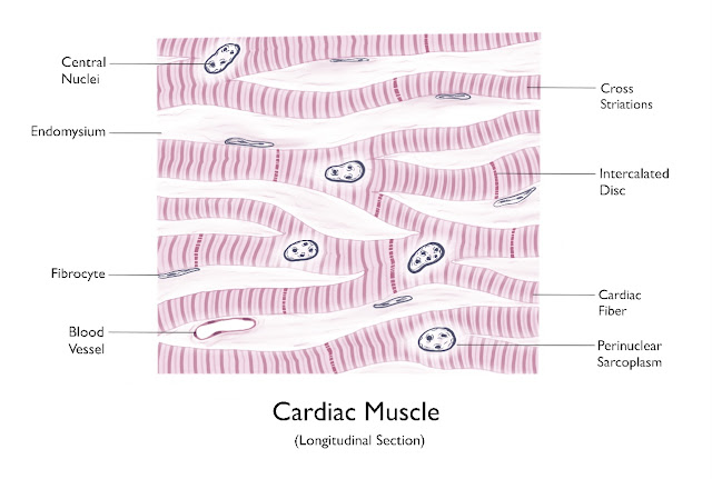
Cardiac Muscle (longitudinal section)

Image
 

Transitional Epithelium

Image
 

Pseudo-stratified Columnar Ciliated Epithelium

Image
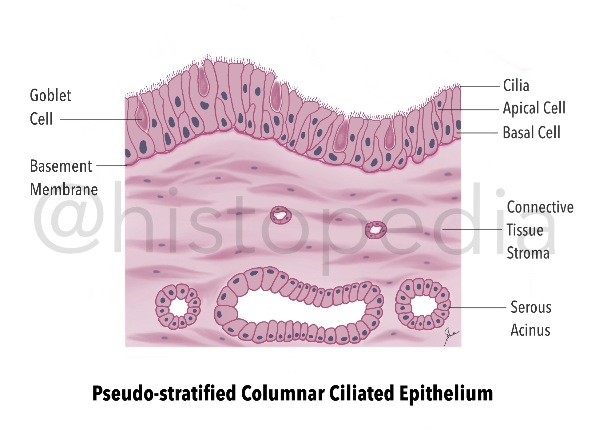
It is the type of epithelium in which cells appear to form several layers, but it is only one layer comprising of cells of different height and shapes and their nuclei are placed in such a fashion which gives pseudo appearance of  multiple layers. Site :  lines Upper Respiratory Passage (trachea & bronchi) Function : Filtration/Humidification of Air in URT Secretion Absorption Other Variants : Non-ciliated pseudo-stratified columnar epithelia (Prostate & Vas deferens) Pseudo-stratified columnar epithelia with   stereo-cilia (Epididymis) Important labels : Cilia Basal cells Apical cells Goblet cells Basement Membrane  

Simple Columnar Epithelium

Image
Single layer of tall and slender cells, with oval nuclei at base attached to basement membrane. Lines the most of the organs of Gastrointestinal tract. Functions: Protection (act as barrier)  Secretory (Stomach) Absorptive (Intestine) In stomach, it exhibits numerous temporary folds. Also extends downwards to form numerous indentations called Gastric Pits .

Simple Squamous Epithelium

Image
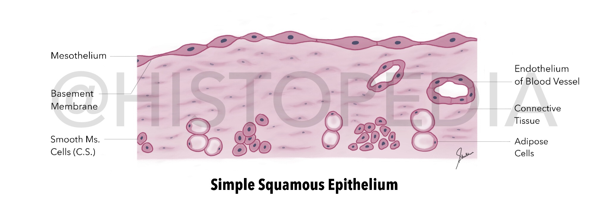
Lines the Body Cavities (Cardiac, Pleural and Peritoneal cavities), the inner surface of blood vessels (endothelium), filtration and diffusion membranes. Primary function is to allow diffusion of small molecules and gases. Important labels: Mesothelium Basement membrane Connective tissue & Smooth Muscle fibres Blood vessels Functions: Allows exchange and transport of fluid/gases Reduces friction Produces transport and lubricating fluids

Skeletal Muscle

Image
 

Connective Tissue Cells

Image