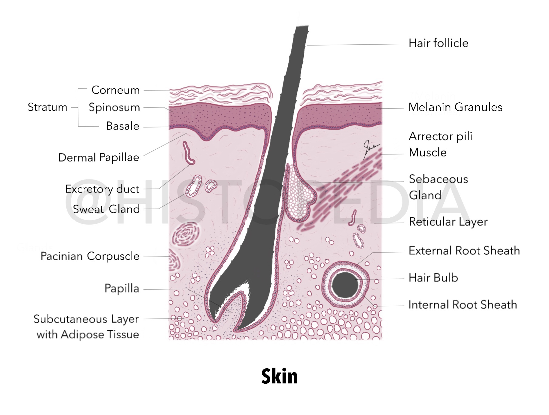
Skin Histology & Thin skin v/s Thick skin

Layers: (top to bottom) Epidermis Stratum Corneum Stratum Spinosum Stratum Basale (with pigmented/melanin granules) Dermis Dermal Papillae (which forms our fingerprints) Reticular layer Subcutaneous layer with Adipose tissue Skeletal muscle fibers (not illustrated here) Hair follicle (numerous, closely packed and oriented at an angle to the surface) Hair bulb Internal root sheath External root sheath Connective tissue Sebaceous Glands : surround each hair follicle, aggregates of clear cells . Arrector pili muscle : smooth ms. aligned at an oblique angle to the hair follicle, it contracts to move hair follicle in more vertical position. Pacinian corpuscles : pressure and vibration sensing receptors. Other labels : sweat glands and blood vessels (omitted here) Imp. Difference between thin skin and thick skin Histology of both types is similar only difference is the thickness of epidermis . As skin of Palm and Soles are constantly exposed to wear/tear/abrasion, thicknes...


