Posts

Parotid Salivary Gland

Image
  Identification Points:  1.   Serous acini   (pyramid shaped secretory cells with round basal nucleus)  2. No Serous Demilune 3.  Striated Ducts Description: It is a large capsulated serous gland, subdivided into lobes and lobules by connective tissue septa located just anterior to the ears. It secretes 25% of total saliva which is serous (watery) in nature.  At higher magnifications small secretory granules are visible in apices of serous acini Flow of Secretions: Serous acini (secretions)  ⤵️ Intercalated ducts   ⤵️ Striated Ducts (lined with Simple Columnar Cells )  ⤵️ Intra-lobular ducts  ⤵️ Interlobular duct ( Pseudo-Stratified Columnar epithelium )  ⤵️ Stensen’s Duct (open near 2nd upper molar) Step-by-step Illustration: File Diagram: How to differentiate between all 3 Salivary Glands?
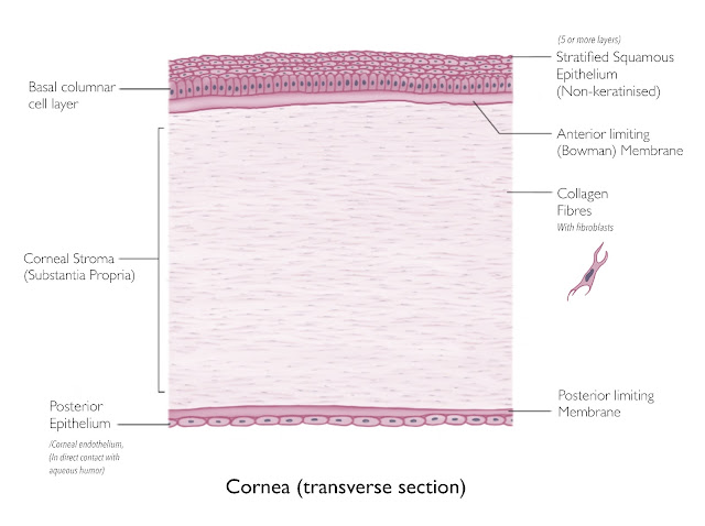
Cornea (transverse section)

Image

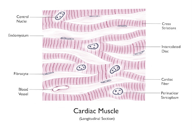
Cardiac Muscle (longitudinal section)

Image
 

Small Intestine: Duodenum (Longitudinal Section)

Image
.  

Tracheal Wall (sectional view)

Image
 

Transitional Epithelium

Image
 

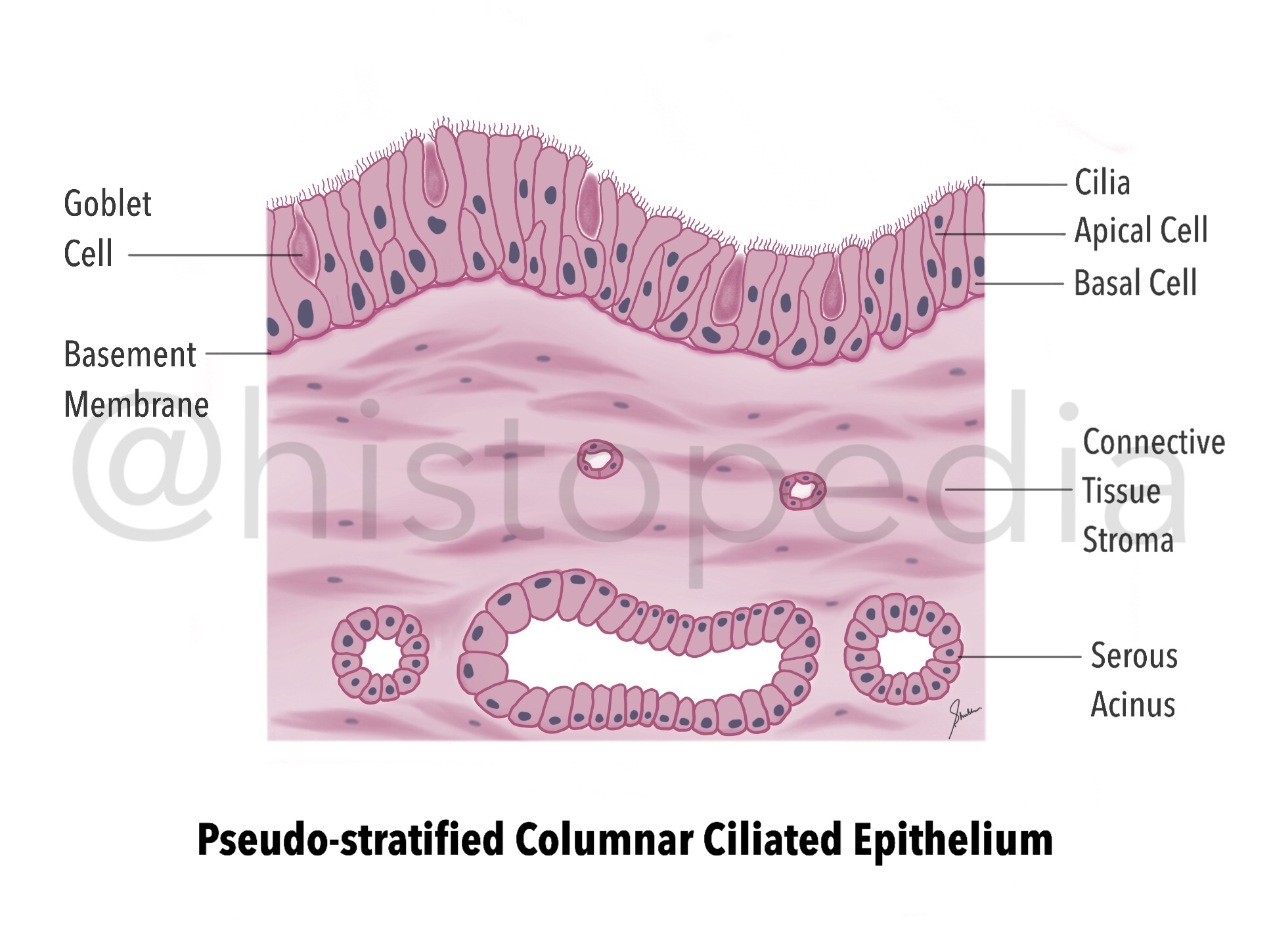
Pseudo-stratified Columnar Ciliated Epithelium

Image
It is the type of epithelium in which cells appear to form several layers, but it is only one layer comprising of cells of different height and shapes and their nuclei are placed in such a fashion which gives pseudo appearance of  multiple layers. Site :  lines Upper Respiratory Passage (trachea & bronchi) Function : Filtration/Humidification of Air in URT Secretion Absorption Other Variants : Non-ciliated pseudo-stratified columnar epithelia (Prostate & Vas deferens) Pseudo-stratified columnar epithelia with   stereo-cilia (Epididymis) Important labels : Cilia Basal cells Apical cells Goblet cells Basement Membrane  

Simple Columnar Epithelium

Image
Single layer of tall and slender cells, with oval nuclei at base attached to basement membrane. Lines the most of the organs of Gastrointestinal tract. Functions: Protection (act as barrier)  Secretory (Stomach) Absorptive (Intestine) In stomach, it exhibits numerous temporary folds. Also extends downwards to form numerous indentations called Gastric Pits .

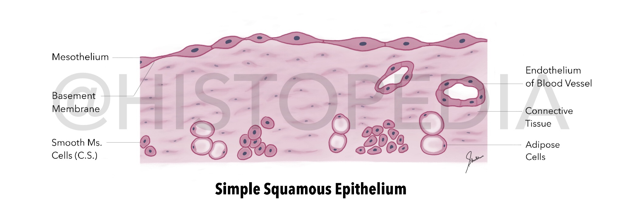
Simple Squamous Epithelium

Image
Lines the Body Cavities (Cardiac, Pleural and Peritoneal cavities), the inner surface of blood vessels (endothelium), filtration and diffusion membranes. Primary function is to allow diffusion of small molecules and gases. Important labels: Mesothelium Basement membrane Connective tissue & Smooth Muscle fibres Blood vessels Functions: Allows exchange and transport of fluid/gases Reduces friction Produces transport and lubricating fluids